
Description
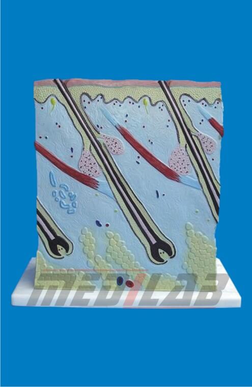
A Section of Skin Model is an essential educational tool used in medical training, biology labs, and dermatology studies to understand the structure and function of human skin. These models provide a cross-sectional view of the skin’s layers and components, helping students and professionals learn about skin anatomy and related medical conditions.
Features of a Section of Skin Model
- Detailed Cross-Section View – Displays the epidermis, dermis, and hypodermis (subcutaneous tissue).
- Color-Coded Layers – Different anatomical regions are highlighted for easy identification.
- Hair Follicle & Gland Representation – Includes hair follicles, sebaceous glands, and sweat glands.
- Blood Vessels & Nerve Endings – Shows capillaries, nerve endings, and sensory receptors.
- Removable Parts – Some models allow for disassembly to study deeper skin structures.
- Durable Material – Made from high-quality PVC or resin for long-lasting use.
- Labeled Structures – Numbered or text-labeled parts for guided learning.
- Enlarged Models – Typically larger than life-size to enhance visibility of microscopic structures.
Types of Section of Skin Models
- Basic Skin Layer Model – Displays the three primary layers: epidermis, dermis, and hypodermis.
- Hair & Gland Model – Includes sebaceous (oil) and sweat glands along with hair follicles.
- Skin Pathology Model – Highlights common skin conditions like acne, eczema, and melanoma.
- Microscopic Skin Model – Shows detailed cellular structures such as melanocytes and keratinocytes.
- 3D Transparent Model – Offers a layered view for better visualization of skin functions.