
Description
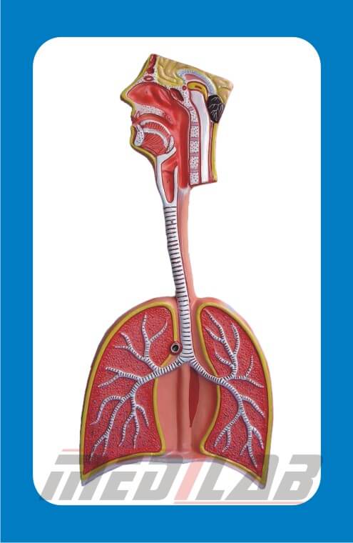
A Human Respiratory System Model is an essential educational tool used in medical schools, biology labs, and healthcare training to study the structure and function of the respiratory system. These models help students visualize how air moves through the body and understand the anatomy of breathing.
Features of a Human Respiratory System Model
- Detailed Anatomical Representation – Includes the nasal cavity, trachea, bronchi, lungs, and diaphragm.
- Color-Coded Structures – Differentiates various parts of the respiratory system for easy identification.
- Removable Parts – Some models allow the lungs to be detached for internal study.
- Durable Material – Made of high-quality PVC or resin for long-term use.
- Labeled Components – Numbered or text-labeled parts for guided learning.
- Life-Size & Enlarged Models – Available in realistic or magnified versions for detailed study.
Types of Human Respiratory System Models
- Basic Respiratory System Model – Shows the complete respiratory tract from the nasal cavity to the lungs.
- Lung Section Model – Provides a cutaway view of lung anatomy, including alveoli.
- Trachea & Bronchi Model – Focuses on the airway structure and bronchial branching.
- Diaphragm & Lung Model – Demonstrates the role of the diaphragm in breathing.
- 3D Transparent Model – Offers a layered view for deeper understanding of air exchange.