
Description
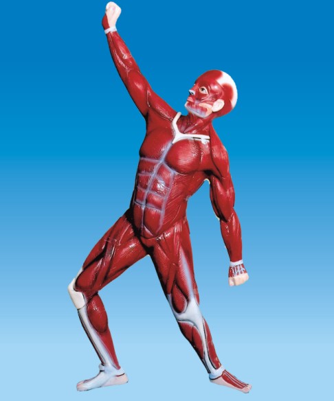
Human Muscular System Model is a valuable educational tool used in medical schools, biology labs, and fitness training to understand the structure and function of human muscles. These models provide a three-dimensional representation of the muscular system, helping students visualize muscle placement, fiber direction, and anatomical relationships.
Features of a Human Muscular System Model
- Detailed Muscle Representation – Displays superficial and deep muscles with realistic texture and structure.
- Removable Parts – Some models allow muscles or muscle groups to be detached for closer examination.
- Material – Made of durable PVC or resin for long-lasting use.
- Life-Size & Scaled Versions – Available in full-scale and smaller sizes for different learning environments.
- Labeled Structures – Many models include numbering or color-coded sections to identify muscles easily.
Types of Human Muscular System Models
- Full-Body Muscular Model – A life-size model showing all major muscles in the human body.
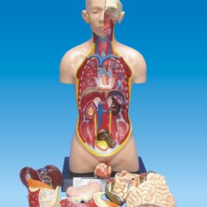
- Torso-Only Muscular Model – Focuses on the muscles of the chest, abdomen, and back.
- Head & Neck Musculature Model – Highlights facial, jaw, and neck muscles.
- Upper & Lower Limb Models – Show detailed musculature of arms or legs separately.
- Layered Models – Provide different layers of muscles for an in-depth study of deep and superficial muscle groups.
Educational Benefits
- Enhances understanding of muscle function and movement.
- Helps medical students, physiotherapists, and fitness professionals visualize muscle interactions.
- Provides a hands-on learning experience compared to flat images in textbooks.
- Supports anatomy education in schools, universities, and medical training centers.