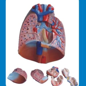

Description
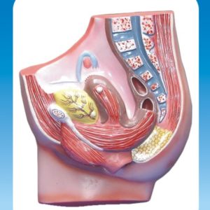
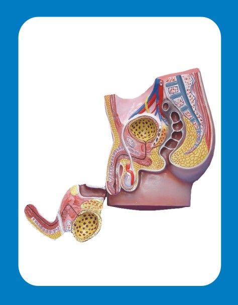
A Human Male Pelvis Model is an essential educational tool for studying the male pelvic anatomy, commonly used in medical schools, anatomy classes, and physiotherapy training. These models provide a three-dimensional representation of the pelvic bones, organs, and surrounding structures, aiding in a better understanding of their functions and relationships.
Features of a Male Pelvis Model
- Detailed Skeletal Structure – Includes pelvic bones, sacrum, coccyx, and acetabulum.
- Reproductive & Urinary Organs – Some models feature the bladder, prostate, seminal vesicles, and rectum.
- Removable Parts – Advanced models allow for the detachment of organs and sections for in-depth study.
- Musculature & Ligaments – Certain models display pelvic floor muscles and ligaments for a complete anatomical view.
- Durable Material – Typically made of high-quality PVC or resin for long-term use.
- Labeled Components – Some versions include numbered or color-coded parts for easier identification.
Types of Male Pelvis Models
- Basic Skeletal Pelvis Model – Includes only the pelvic bones without soft tissues.
- Male Pelvis with Organs – Features internal structures like the prostate, bladder, and rectum.
- Pelvic Floor Musculature Model – Highlights the muscles supporting the pelvic organs.
- Cross-Section Pelvis Model – Provides a detailed sectional view of internal structures.
- 3D Printed or Transparent Models – Offer a unique visualization of internal layers and functions.
Educational Benefits
- Enhances understanding of male reproductive and urinary systems.
- Aids medical students, urologists, and physiotherapists in anatomy training.
- Supports study in conditions related to the prostate, bladder, and pelvic floor dysfunction.
- Provides a hands-on, visual learning experience for better retention.