
Description
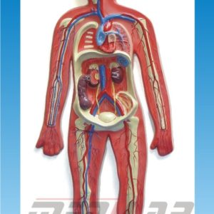
A Human Digestive System Model is an essential educational tool used in medical schools, biology labs, and healthcare training to study the anatomy and function of the digestive system. These models help students visualize the structure and spatial arrangement of digestive organs, aiding in a better understanding of digestion, absorption, and gastrointestinal health.
Features of a Human Digestive System Model
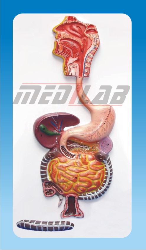
- Detailed Anatomical Representation – Includes major organs such as the esophagus, stomach, liver, pancreas, intestines, and rectum.
- Removable Parts – Some models allow for the detachment of organs to examine internal structures.
- Labeled Components – Numbered or color-coded sections for easy identification.
- Durable Material – Made of PVC, resin, or rubber for long-term use.
- Life-Size & Miniature Versions – Available in full-scale for realistic study or smaller models for compact learning.
- Wall-Mounted or 3D Free-Standing – Some models are mounted for classroom display, while others are standalone for hands-on study.
Types of Human Digestive System Models
- Basic Digestive System Model – Shows a simplified representation of the entire system.
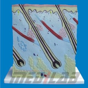
- Detailed Sectional Model – Provides a closer look at internal organ structures, such as stomach layers or intestinal villi.
- Digestive Tract Model – Focuses on the pathway from the esophagus to the rectum.
- Liver, Stomach & Pancreas Model – Highlights the role of accessory organs in digestion.
- 3D Transparent Model – Offers a layered view for understanding digestion mechanics.