
Description
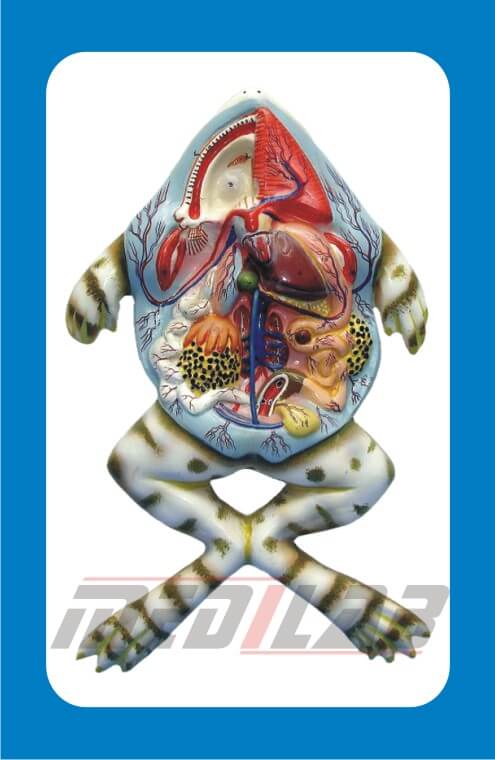
Frog Dissection Model is an essential educational tool used in biology and anatomy studies to help students understand the internal and external anatomy of a frog. It provides a detailed representation of the frog’s organs, making it an excellent alternative to real dissections in classrooms.
Features of a Frog Dissection Model
- Lifelike Anatomical Structure – Accurately represents the internal and external organs of a frog.
- Color-Coded Organs – Different colors help distinguish the heart, lungs, liver, stomach, intestines, and other organs.
- Detachable Parts – Some models have removable organs for interactive learning.
- Durable Material – Made from PVC, plastic, or resin for long-term use.
- Realistic Texture & Detailing – Mimics the actual look of a dissected frog.
- Mounted or Free-Standing Design – Can be placed on a base or used as a handheld model.
- Labeled Structures – Numbered or text-labeled parts for easy identification.
Types of Frog Dissection Models
- Basic Frog Dissection Model – Displays essential internal organs.
- 3D Layered Model – Shows multiple layers of dissection for in-depth study.
- Removable Organ Model – Features detachable parts for hands-on learning.
- Transparent Frog Model – Provides a clear view of the internal structure without opening the frog.
- Magnetic Frog Model – Includes removable magnetic components for interactive education